
Laboratory of Biologically Active Compounds"Shared-Use Centre"
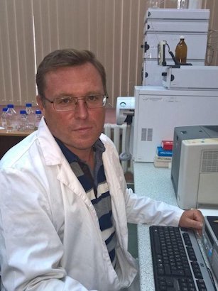
Head of the Centre
Konovets Ihor Mykolaiovych
Senior Researcher, Cand. Biol. Sci. 📍 Kyiv, Volodymyra Ivasiuka Ave., 12; room 216 📞 38 (044) 419-39-81 📠 38 (044) 418-22-32
Centre Regulations
Establishment of the Centre of the Institute of Hydrobiology, NAS of Ukraine
The Hydroecological Analytical Shared-Use Centre for scientific instruments/equipment of the Institute of Hydrobiology of the NAS of Ukraine was established with the aim of the most rational use of unique and costly scientific instruments/equipment of domestic or foreign manufacture, which it is impossible or impractical for each scientific institution to acquire from centralised funds, joint funds of groups of institutions and organisations, or other NAS of Ukraine funding sources.
Structure, Main Objectives and Organisation of Work
2.1. The Hydroecological Analytical Centre is a component of the Scientific and Technical Shared-Use Centre for scientific instruments and equipment in the scientific (scientific-technical) direction, a property of the National Academy of Sciences of Ukraine.
2.3. The Hydroecological Analytical Centre of the Institute of Hydrobiology of the NAS of Ukraine is a scientific subdivision directly subordinated to the Directorate of the Institute without the status of a legal entity.
2.4. The main objective of the HAC of the Institute of Hydrobiology is to provide NAS of Ukraine scientists with the opportunity to conduct research on state-of-the-art scientific instruments/equipment maintained by qualified personnel capable of keeping the equipment in high-quality working condition and providing advisory services.
Service Provision Procedure
3.1. Information about the HAC of the Institute of Hydrobiology of the NAS of Ukraine is available on the Institute's website and on the website of the Presidium of the NAS of Ukraine.
3.2. Scientific institutions and organisations of the NAS of Ukraine that need to conduct research using the HAC's scientific equipment submit in writing to the Bureau of the Division of General Biology and the HAC IHB their requests for the number of hours, duration, and types of research — twice a year, before 15 January and before 15 June.
3.3. The Bureau of the Division of General Biology of the NAS of Ukraine, upon the submission of the Director of the Institute of Hydrobiology of the NAS of Ukraine, approves the overall distribution among clients of the working time allocated for shared use of the Centre's scientific equipment.
3.5. The HAC of the Institute of Hydrobiology of the NAS of Ukraine establishes the following schedule of instrument use during a single-shift working day (8 hours):
3.6. Paid services for the use of HAC scientific equipment are provided in accordance with the current legislation of Ukraine.
Scientific Equipment
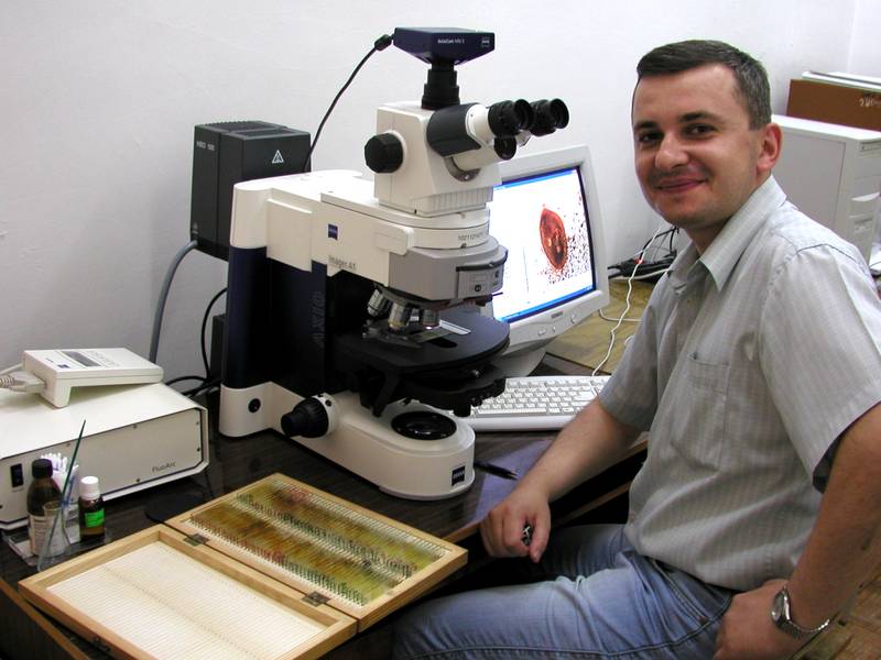
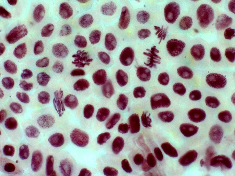
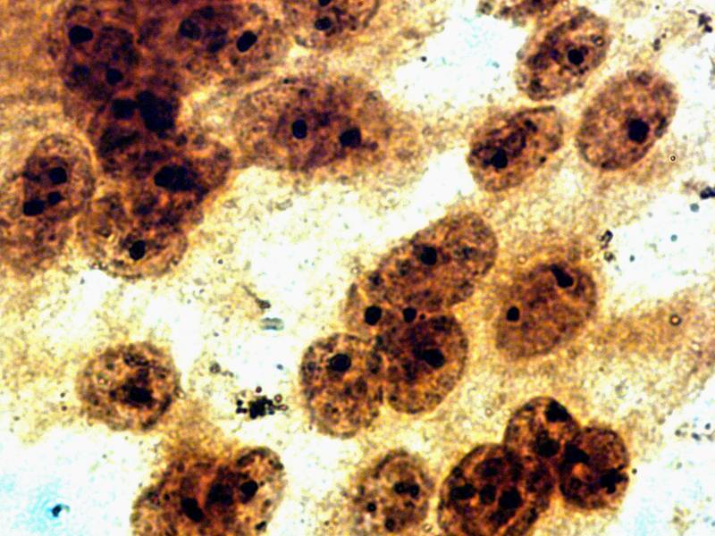
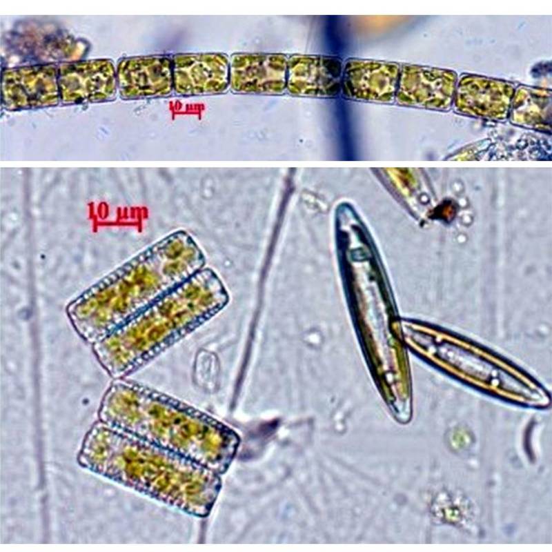
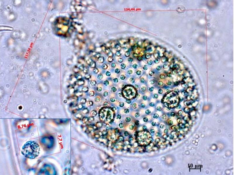
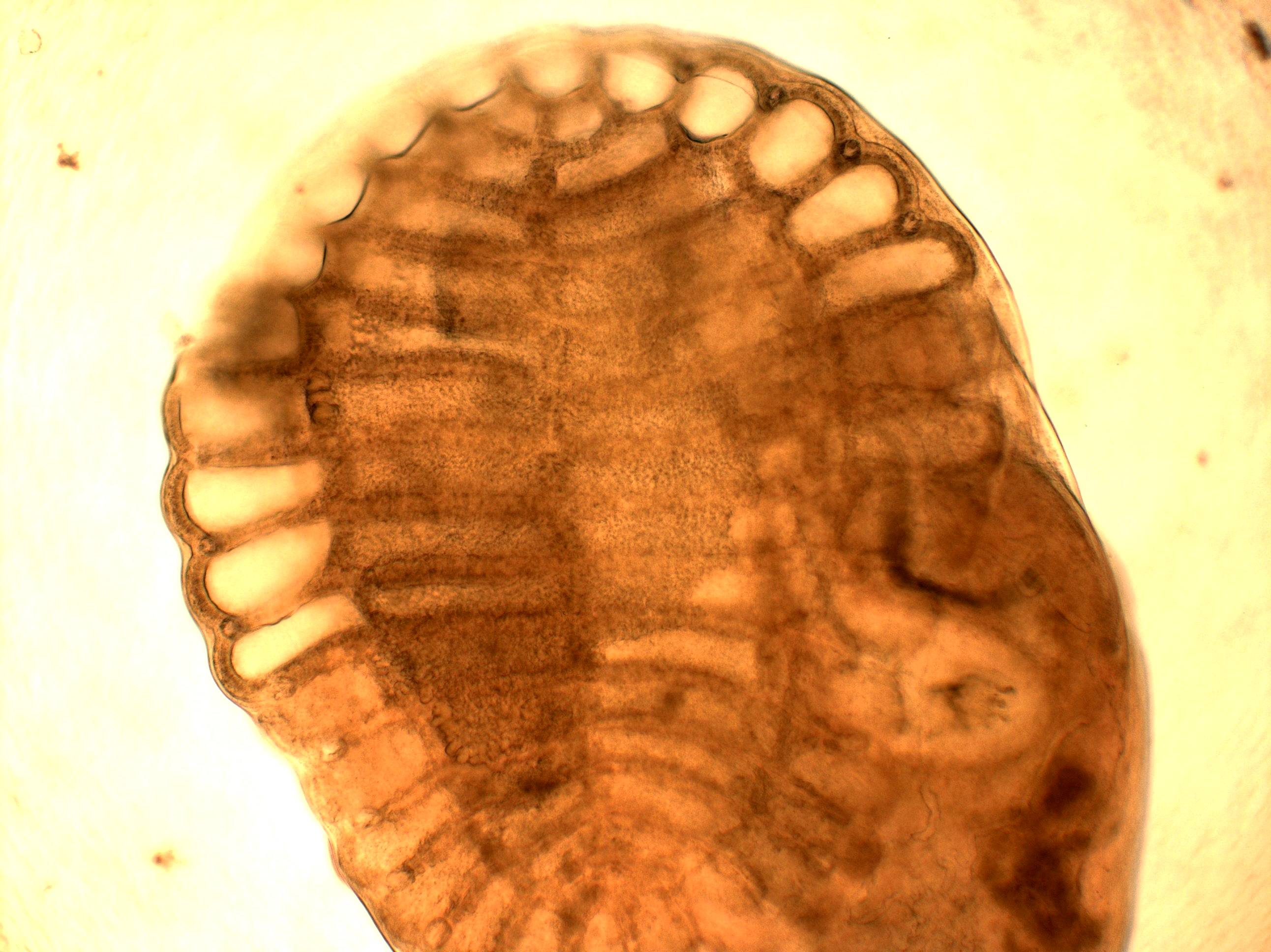
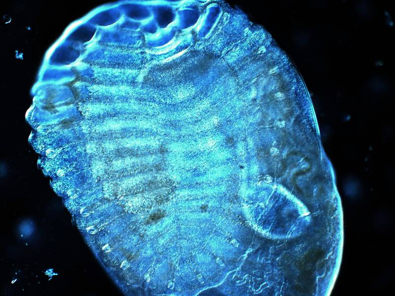
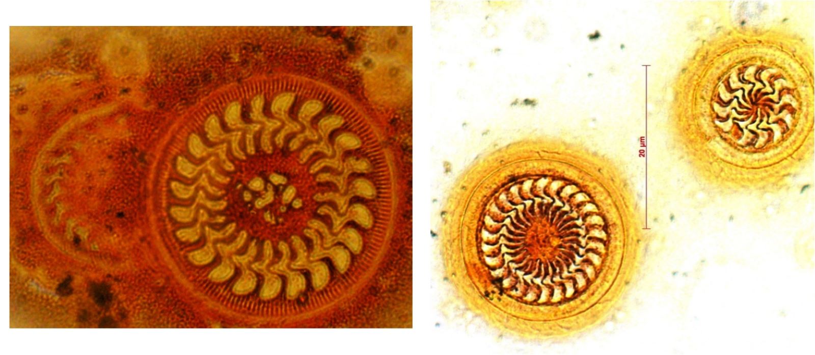
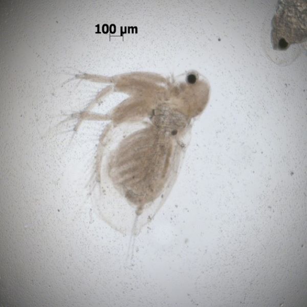
Research Microscope Axio Imager A1 "Carl Zeiss" (Germany)
Application: a universal and applied microscope for use in biology and medicine. Complies with international standard ISO 9000.
Microscopy methods:
- In transmitted light — brightfield and differential interference contrast.
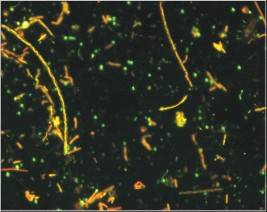
- In reflected light — epifluorescence.
Objectives with magnification/numerical aperture: 2.5×/0.085; 5×/0.15; 10×/0.30; 20×/0.50; 40×/0.75; 63×/0.85; 100×/1.30. Optical correction of objectives — apochromatic.
Filter sets for epifluorescence:
- Set No. 15 — excitation BP 546/12, beamsplitter FT 580, emission LP 590
- Set No. 44 — excitation BP 475/40, beamsplitter FT 500, emission LP 530/50
- Set No. 49 — excitation G 365, beamsplitter FT 395, emission BP 445/50
Imaging system: AxioCam 506 color digital camera (6 MP) with ZEN 2.6 software.
Automated Liquid Chromatograph-Mass Spectrometer Agilent 1200 SL/DAD/FD/MSD 6130
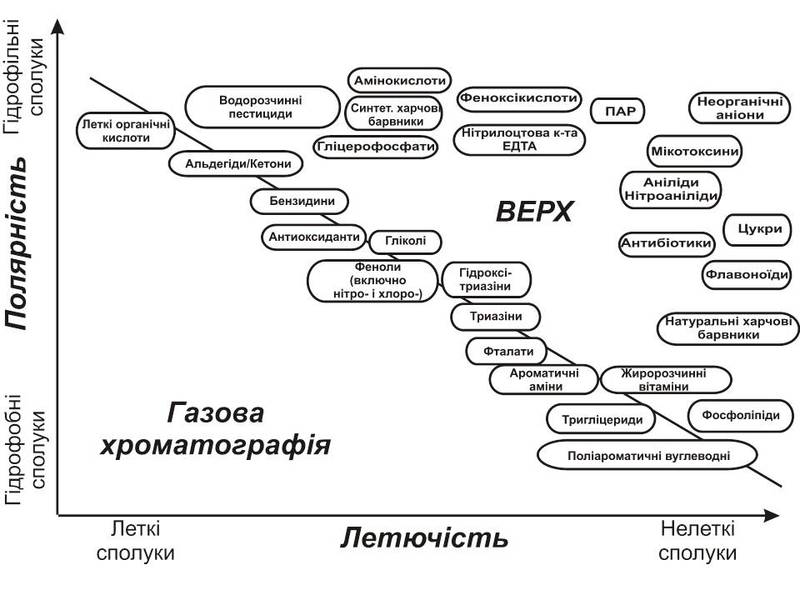
Application: qualitative and quantitative determination of polar non-volatile thermolabile and thermostable organic compounds — pharmaceuticals, physiologically active substances, water-soluble pesticides, polyaromatic hydrocarbons, food additives, and other toxicants and compounds of organic origin — in matrices of varying complexity.
Detectors:
- Diode-array detector (DAD) — multi-wavelength analysis and spectral recording (190–950 nm)
- Fluorescence detector (FD) — multi-wavelength detection and rapid fluorescence spectrometry
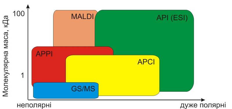
- Single-quadrupole mass-selective detector (MSD 6130) with electrospray (API) and chemical ionisation (APCI) sources
Illustrations
Selected Publications
- Савлучинська М.О., Коновець І.М., Арсан О.М., Мардаревич М.Г. Особливості енергозабезпечення процесів адаптації риб до різних концентрацій фіпронілу та диметоату у водному середовищі // Наук. зап. Терноп. нац. пед. ун-ту. Сер. Біол. – 2017. – T. 69, №2. – С. 70–75.
- Rodriguez R., Vakulenko O., Ralchenko S., Konovets I. et al. Quantification of S-adenosyl methionine and S-adenosyl-homocysteine in human placenta and placental explants under homocysteine treatment // International Journal of Mass Spectrometry. – 2017. – Vol. 421. – P. 279–284. DOI: 10.1016/j.ijms.2017.08.002.
- Usenko O.M., Konovets I.N., Tarashchuk O.S., Gorbunova Z.N. Phenolcarbonic acids of the submerged aquatic plants and their effect on phytoepiphyton structure // Hydrobiological Journal. – 2019. – Vol. 55, N 6. – P. 55–64. DOI: 10.1615/HydrobJ.v55.i6.50.
- Ho K.T., Konovets I.M., Terletskaya A.V. et al. Contaminants, mutagenicity and toxicity in the surface waters of Kyiv, Ukraine // Marine Pollution Bulletin. – 2020. – Vol. 155. – P. 111153. DOI: 10.1016/j.marpolbul.2020.111153.
- Rodriguez R., Konovets I., Ralchenko S. et al. A low-cost mass spectrometry-based approach for quantifying purines in placental explants // International Journal of Mass Spectrometry. – 2021. – Vol. 460. – Art. 116490. DOI: 10.1016/j.ijms.2020.116490.